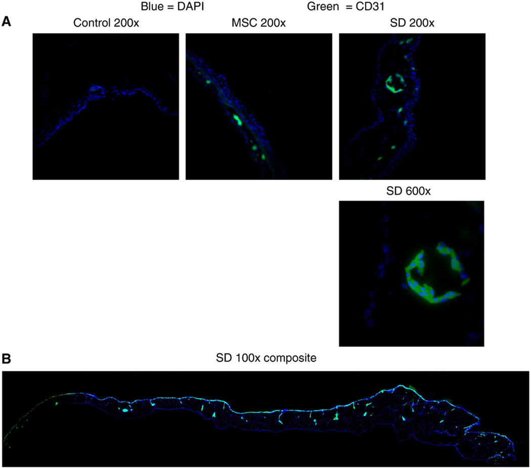
Figure 7.
SD-MSCs express CD31 in vivo. (A) Representative immunofluorescent images of chick CAMs after growth with grBME-containing MSCs, SD-MSCs on Day 30 of serum deprivation, or vehicle control. Merged color images are displayed for SD-MSCs (SD), MSCs, and a vehicle control. (B) Composite image of chick CAM grown with SD-MSCs is also shown. Multiple images were taken at a magnification of 100× and then combined to create an image displaying the entire CAM. Green represents staining with a human specific CD31 antibody and blue represents DAPI staining.

