Fig. 1.
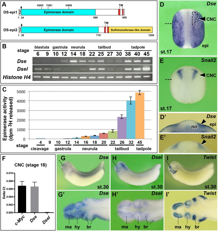
Expression and activity of the two dermatan sulfate epimerases in Xenopus embryos. (A) Protein structures. Xenopus DS-epi1 and DS-epi2 contain cleavable signal peptides (SP, arrows), an epimerase domain and two transmembrane (TM) domains. In DS-epi1, the catalytic residues His205, Tyr261 and His450 are indicated, which are also conserved in DS-epi2. DS-epi2 contains an additional sulfotransferase-like domain. (B) RT-PCR analysis of Dse and Dsel. Histone H4 is used as the loading control. A minimum of two experiments (n≥2) was performed with three independent biological samples. (C) DS epimerase activity in early Xenopus embryos. Results are mean±s.d. from triplicates (two independent experiments). (D-E′) Whole-mount in situ hybridization of neurula embryos in the dorsal view (D,E) and in transversal section (D′,E′). The arrowheads label the pre-migratory CNC cells. The region enclosed by the dashed line demarcates the Snail2+ CNC embedded in the Dse expression domain. (F) qPCR analysis in CNC explants at stage 18. c-Myc was used as a CNC cell marker. Note that Dse but not Dsel mRNA is detected. Results are mean±s.d. from triplicates (n=4 biological replicates). (G-I′) Tailbud embryos in the lateral view (G-I) and horizontal section (G′-I′). Note that Dse and Dsel overlap with Twist expression in migrating CNC cells. Section planes are indicated with dashed straight lines. br, branchial arch segments; epi, epidermis; hy, hyoid segment; ma, mandibular segment; no, notochord.

