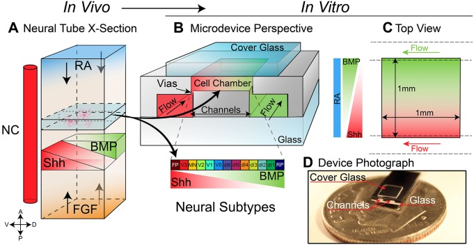
Fig. 1.
Graphical overview of the microfluidic reconstruction of the neural tube. (A,B) Schematic of a neural tube highlighting the 100 µm ‘slice’ recreated by the microfluidic device. Four primary signals (RA, SHH, BMP and FGF) are responsible for patterning the bulk of the neural tube (NC, notochord) (A). The SHH gradient, which is responsible for directing the differentiation of ventral neural progenitors into discrete domains of neurons, is recreated inside the cell culture chamber of the microdevice (B). Flow channels running under the cell chamber supply nutrients as well as desired guidance molecules to the cells in the culture chamber. Morphogen concentration gradients are established across the chamber using the vias in a standard source/sink configuration with the walls of the chamber acting as reflective boundaries. (C) A top (rotated) view of B, as seen through the cover glass. (D) A photograph of the microdevice sitting on top of a dime indicates the scale of the device. FP, floor plate; RP, roof plate.

