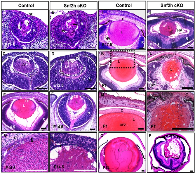
Fig. 2.
Snf2h is necessary for mouse lens development. (A,B) Lens vesicle is separated from the surface ectoderm in the Snf2h cKO at E11.5. Note that a number of cells at the posterior of the lens vesicle (LV) in the Snf2h cKO already appeared to elongate (arrow in B). (C,D) Primary lens fiber cells are elongated within the lens vesicle at E12.5. The prominent hyaloid vasculature (HV) occupies the space between the lens (L), cornea (C) and retina (R) in the Snf2h cKO eyes. (E-H) At E14.5, lens fiber cells in the Snf2h cKO were unable to form the bow region/transitional zone and the fiber-like morphology of these cells deteriorated at the anterior of the lens. (I,J) The E17.5 Snf2h cKO shows a series of defects in lens, cornea, iris (I) and retina. (K-N) The newborn Snf2h mutant lens does not form the presumptive organelle free zone (OFZ). The boxed region in K is magnified in M. (O,P) Profound ocular defects in Snf2h cKO are found at P14, including the absence of lens anterior chamber, lens vacuoles, and disorganization of the lens fiber cells. LE, lens epithelium; LF, lens fibers; SE, surface ectoderm. Scale bars: 100 μm.

