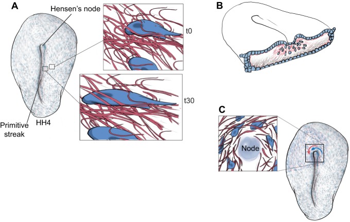
Fig. 1.
Composite tissue (cells plus ECM) motion during early embryogenesis. (A) ECM motion is present in avian embryos as early as primitive streak formation. Time-lapse recordings of cells (blue) and ECM fibers (red) show both embryonic tissue components moving as a composite [compare insets at time (t) 0 and 30 min later], along with concomitant cell shape changes. (B) During gastrulation, ECM fibers (pink) move through the primitive streak (ingress) along with prospective mesendodermal cells (blue). See also Movie 1. (C) A third example of ECM fiber motion occurs near Hensen's node (the ‘organizer’ in birds). Nodal cells, adjacent epiblastic cells and ECM fibers move in unison (blue and red arrows). The organizer tissue undergoes rotational motion that determines left-right asymmetry with respect to the head-to-tail axis. Only counterclockwise motion is illustrated for simplicity; however, clockwise rotation of more remote epiblastic cells occurs in the peri-nodal region (see Cui et al., 2009b).

