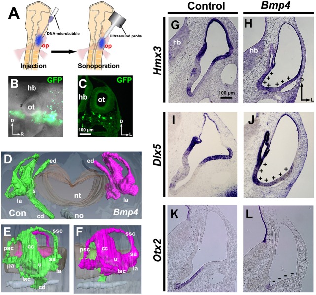
Fig. 1.
Bmp4 overexpression expands Hmx3 and Dlx5 expression ventrally, attenuates Otx2 expression and blocks formation of the cochlea. (A-C) Transfection scheme. (A) Diagram showing sonoporation targeted to transfect head mesenchymal cells ventral to the otic placode. (B) Whole-mount view 12-18 h after sonoporation of Gfp. (C) Transverse section through another transfected chicken embryo. (D-F) Reconstruction (D, frontal view; E,F, lateral views) of serial transverse sections of inner ears (HH stage 30) collected after sonoporation of Bmp4. Green, control (Con); magenta, Bmp4 overexpression. Paint fills, n=3; paraffin serial sections, n=5; reconstruction, n=1. (G-L) In situ hybridization of transverse sections of otocysts collected after transfection when embryos reached HH stages 24-25. (G,H) Hmx3 expression. +, region of expanded Hmx3 expression. (I,J) Dlx5 expression. +, region of expanded Dlx5 expression. (K,L) Otx2 expression. −, region of reduced Otx2 expression. n=6 (G,H,K,L) or n=9 (I,J) control and experimental otocysts. The right-left axis of histological images and reconstructions are oriented here and subsequently to align with the experimental schema, simplifying data interpretation. D, dorsal; L, lateral; R, rostral; cc, common crus; cd, cochlear duct; ed, endolymphatic duct; hb, hindbrain; la, lateral ampulla; lsc, lateral semicircular canal; no, notochord; nt, neural tube; op, otic placode; ot, otocyst; pa, posterior ampulla; psc, posterior semicircular canal; s, saccule; sa, superior ampulla; ssc, superior semicircular canal; u, utricle.

