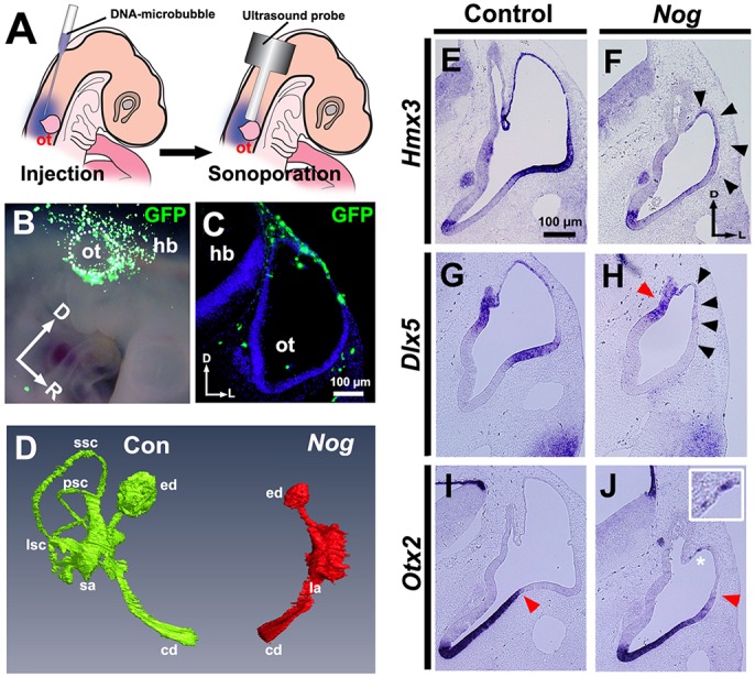
Fig. 2.

Nog overexpression attenuates Hmx3 and Dlx5 expression, expands Otx2 expression dorsally and blocks formation of the semicircular canals. (A-C) Transfection scheme. (A) Diagram showing sonoporation targeted to transfect head mesenchymal cells overlying the dorsal otocyst. (B) Whole-mount view 12-18 h after sonoporation of Gfp. The borders of the otocyst are indicated by white dots. (C) Transverse section through another transfected embryo. (D) Frontal view of a reconstruction of serial transverse sections of inner ears at HH stage 35 collected after sonoporation of Nog. Green, control (Con); red, Nog overexpression. Paint fills, n=3; paraffin serial sections, n=3; reconstruction, n=1. (E-J) In situ hybridization of transverse sections of otocysts collected after transfection when embryos reached HH stages 24-25. (E,F) Hmx3 expression. Arrowheads indicate region of reduced Hmx3 expression. (G,H) Dlx5 expression. Black arrowheads indicate region of abolished Dlx5 expression; red arrowhead indicates region (the developing endolymphatic duct) of persisting Dlx5 expression. (I,J) Otx2 expression. Asterisk indicates ectopic expression of Otx2 (enlarged in inset); red arrowhead indicates the dorsolateral limit of Otx2 expression, which is expanded further dorsally after Nog overexpression. (E-J) n=6 control and experimental otocysts. Abbreviations as in Fig. 1.
