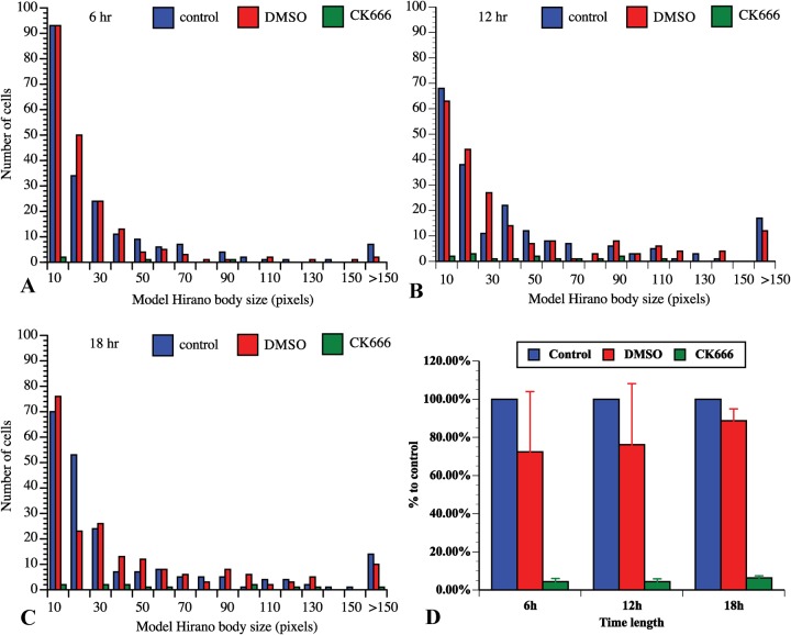
Fig. 4.
CK666 affects the distribution of model Hirano body size and number of cells that generate model Hirano bodies. The size distribution of model Hirano bodies was measured after 6 h (A), 12 h (B) or 18 h (C) of incubation with CK666 in media (green) after removal of folic acid to induce expression of E60K-GFP, compared with cells incubated with DMSO plus media (red) or media only (blue) under the same condition to induce model Hirano bodies. The distributions of the model Hirano body size were significantly different (P<0.01 at 6 h, P<0.001 at 12 h and at 18 h). The proportions of cells that generated model Hirano bodies with or without CK666 were also counted, and compared with the control (D). The number of cells that produced model Hirano bodies were significantly reduced when incubated in the presence CK666 (P<0.001 for all three time points, data represented as means±S.D.).

