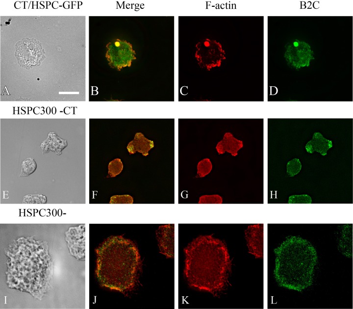
Fig. 6.
Localization of HSPC300 and F-actin in Dictyostelium cells expressing model Hirano bodies. (A-D) Cells expressing CT were transformed with plasmid to express HSPC300-GFP, and were observed by DIC microscopy (A), and by fluorescence microscopy using either TRITC-phalloidin (C) or GFP (D). HSPC300 is enriched in model Hirano bodies (B,D). (E-L) HSPC300 is required for model Hirano body formation. HSPC300− transformed with CT (E-H), and control HSPC300− (I-L), were examined by DIC microscopy (E,I), and by fluorescence microscopy using TRITC-phalloidin (G,K) or B2C that recognizes CT (H,L). No model Hirano bodies were observed. Scale bar=10 µm.

