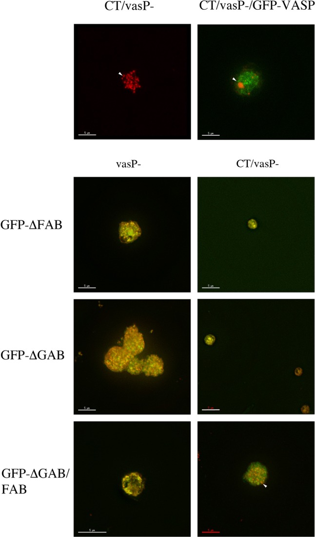
Fig. 8.

Localization of VASP and mutant forms of VASP and F-actin in Dictyostelium cells transformed with CT to induce model Hirano bodies. vasP− were transformed with plasmid to express GFP-VASP, GFP-ΔGAB, GFP-ΔFAB, GFP-ΔGAB/FAB and in the presence or absence of CT to induce model Hirano bodies. The cells were observed by fluorescence microscopy using either TRITC-phalloidin or GFP. Except for CT/vasP–, the merged TRITC-phalloidin and GFP images are shown. When CT is expressed in vasP−, punctate small actin foci were formed. Model Hirano bodies were formed when CT/vasP− was rescued with full length GFP-VASP. Small foci containing either actin, VASP, or both were observed when either GFP-ΔGAB or GFP-ΔFAB VASP was expressed in vasP−. In the presence of CT and either GFP-ΔGAB or GFP-ΔFAB VASP, no large model Hirano bodies were formed, only small foci. When CT is expressed in GFPΔGAB/FAB vasP−, punctate actin foci that resembled those formed in vasP– cells were observed. VASP and its ability to bind actin is required either for F-actin foci to coalesce or elongate from F-actin foci to form model Hirano bodies. Arrowheads indicate either actin foci or model Hirano bodies. Scale bar=5 µm.
