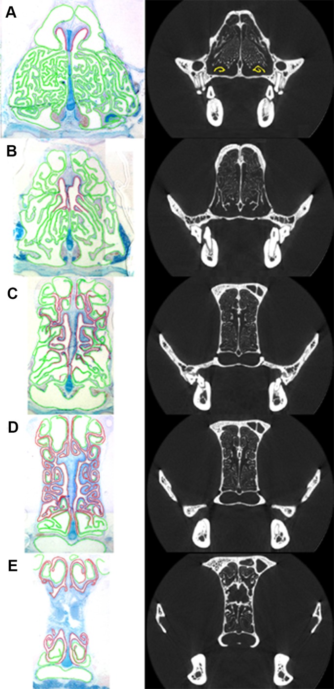
Fig. 2.

Coronal view of histological sections (left) of the turbinals of cat 6559 with olfactory epithelium (OE; red) and non-sensory epithelium (green), and coronal CT scans (right) corresponding to approximately similar locations in the skull. CT scan A has the maxilloturbinals colored in yellow, showing minimal maxilloturbinal presence at this location. See Fig. 1 for location of scans A–E within the skull.
