Fig. 5.
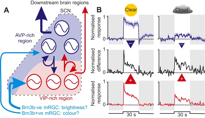
SCN network organisation and possible implications of sensory processing. (A) A proposed model of SCN organisation comprising reciprocally (and locally) coupled vasoactive intestinal polypeptide (VIP)- and arginine-vasopressin (AVP)-rich regions (red and blue, respectively). The VIP-cell region receives dense retinal input (thick light blue arrow) and contains weak oscillators (represented by shallow waves) that are easily reset by external stimuli. The AVP-cell region receives less retinal innervation (thin light blue arrow), but contains more robust oscillators (deeper waves) and provides more extensive innervation of downstream brain regions (compare thick dark blue arrow versus thin red arrow). Retinal input to the two regions derives from genetically distinct mRGC subtypes, which are either Brn3b positive or negative (Brn3b+ve or −ve, respectively), proposed here to reflect cells providing chromatic versus achromatic input, respectively. (B) The normalised (relative to maximal firing) activity of blue-ON cells (top) and achromatic cells (bottom) to light steps replicating the colour and brightness of natural daylight (solar elevation=+3 deg) on clear (left panels) or cloudy days (right panels). Red and blue triangles represent the approximate strength of proposed excitatory (+) and inhibitory (−) contributions to phase resetting. Middle panels in B represent the difference between achromatic and blue-ON cell activity (weighted according to the twofold greater density of achromatic cells). Note that, because blue-ON cell activity is much more strongly suppressed under cloudy days than that of achromatic cells, the difference in activity of the two populations for a fixed solar elevation is broadly similar regardless of weather conditions. Periods of light are represented by white backgrounds and periods of darkness are represented by grey backgrounds. Data in B are derived from Walmsley et al. (2015).

