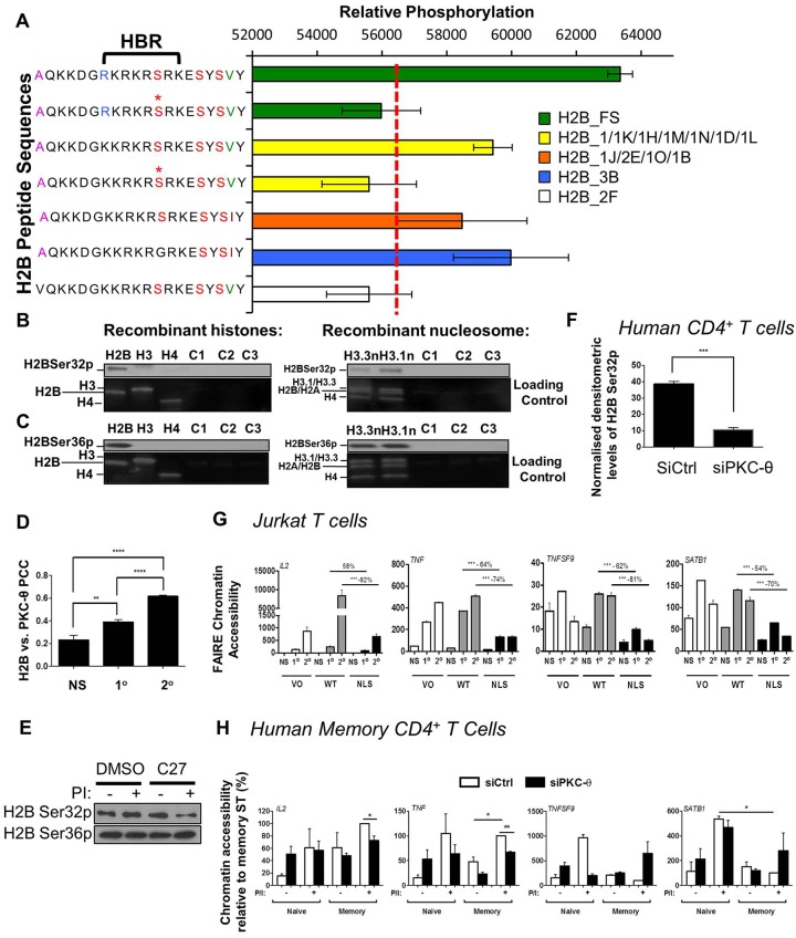
Fig. 7.
Identification of phosphorylated residues on histone H2B. (A) PKC-θ-mediated phosphorylation signals on H2B:21 (residues 21–40 derived from H2B) were detected by PKC-θ microarray profiling. The mean phosphorylation is shown (±s.d.). * denotes a pre-phosphorylated serine and the dotted red line is the background threshold (57500). The location of the histone H2B repression domain (HBR) is marked. (B) PKC-θ phosphorylates H2B Ser32. An in vitro kinase assay was performed by incubating active PKC-θ with either recombinant histones H2B, H3 or H4 or recombinant nucleosomes containing H3.1 or H3.3. Phosphorylated proteins were resolved by SDS-PAGE followed by western blotting for H2B phosphorylation. Negative controls include C1 (no ATP addition), C2 (no PKC addition), and C3 (incubation with PKC-μ). A representative blot of three experiments is shown. (C) PKC-θ phosphorylates H2B Ser36, as assessed by the method shown in B. (D) The Pearson's colocalization coefficient (PCC) was calculated for the fluorescent signal of H2B and PKC-θ as measured by confocal laser scanning microscopy in non-stimulated (NS) Jurkat T cells, and cells after primary (1°) and secondary (2°) stimulations (mean±s.e.m., n=20). **P≤0.01, ****P≤0.0001 (Mann–Whitney test). (E) Representative immunoblot of phosphorylated H2B Ser32 and Ser36 in DMSO and 1 μM C27-treated Jurkat T cells with or without PMA and Ca2+ ionophore (PI) (n=3). (F) Normalized H2B Ser32p densitometry is shown for human CD4+ T cells treated with either the control siRNA (siCtrl) or PKC-θ siRNA (siPKC-θ) (mean±s.e.m., n=3 individuals). ***P≤0.001 (two-tailed Student's t-test). (G) FAIRE chromatin accessibility shown for IL2 and TNF in in non-stimulated (NS) Jurkat T cells, and cells after primary (1°) and secondary (2°) stimulations transfected with vector only (VO), wild-type PKC-θ plasmid (WT) or cytoplasmic-restricted PKC-θ mutant (NLS) plasmids. FAIRE chromatin accessibility is normalized to results for GAPDH (mean±s.e.m., n=3). ***P≤0.001 (two-way ANOVA). (H) FAIRE chromatin accessibility shown for IL2 and other promoters in naïve and memory CD4+ T cells treated with either the control (siCtrl) or the PKC-θ siRNA (siPKC-θ) with or without PMA and Ca2+ ionophore (P/I). FAIRE chromatin accessibility is normalized to GAPDH and expressed as a percentage relative to the stimulated (ST) memory CD4+ T cells treated with the control siRNA (siCtrl) (mean±s.e.m., n=3). *P≤0.05, **P<0.01 (unpaired two-tailed Student's t-test).

