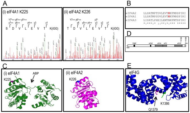
Fig. 5.
eIF4A1 and eIF4A2 are sumoylated on K225 and K226, respectively. (A) Mass spectra of sumoylation products. High-molecular-mass species from an in vitro sumoylation assay were excised from SDS-PAGE gels and analysed by LC-MS/MS. (B) Sequence alignment of human eIF4A1, eIF4A2 and eIF4A3. Sumoylation sites are indicated in red. (C) Position of sumoylation sites on eIF4A1 (i) and eIF4A2 (ii). Pymol-derived figure indicating sumoylation sites on human eIF4A1 (PDB ID 2ZU6) and eIF4A2 (PDB ID 3BOR). ABP, ATP-binding pocket. (D) Schematic indicating organisation of interacting motifs in eIF4G. (E) Positions of sumoylation site (K1386, green) and Q1379 (magenta) in the C-terminal fragment of eIF4G (PDB IB 1UG3).

