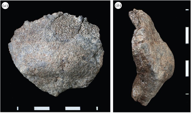
Figure 4.
The partial occipital bone KNM-ER 2598 from the upper Burgi Member of the Koobi Fora Formation (a, posterior view; b, left lateral view). Thick bone (seen in cross section), a well-developed occipital torus (especially centrally), and the low position of the internal occipital protuberance relative to inion affiliate this specimen with the Homo erectus morphological pattern [61]. The widely divergent limbs of the lambdodial suture are also characteristic of many African and Asian H. erectus crania. Photos courtesy of Fred Spoor (scale bar, 1 cm). (Online version in colour.)

