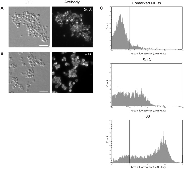
Fig 5. Comparison of the anti-SctA and H36 antibodies as MLB markers.
Immunofluorescence analyses of purified MLBs using the (A) anti-SctA and (B) H36 antibodies. SctA labeling was not uniform and the protein appeared to be concentrated in aggregates in the MLBs. H36 labeling was only seen on the surface of MLBs, creating a ring-like appearance. The purified MLBs were deposited on glass slides and were processed for immunofluorescence using the anti-SctA or H36 antibodies. (C) Flow cytometric analysis of purified MLBs labeled with the anti-SctA or H36 antibody. Approximately 40% of the MLBs were labeled with the anti-SctA antibody while 60–70% were labeled with the H36 antibody. The fluorescence of the labeled MLBs was much more intense with the H36 antibody than with the anti-SctA antibody. Scale bar: A and B = 5 μm.

