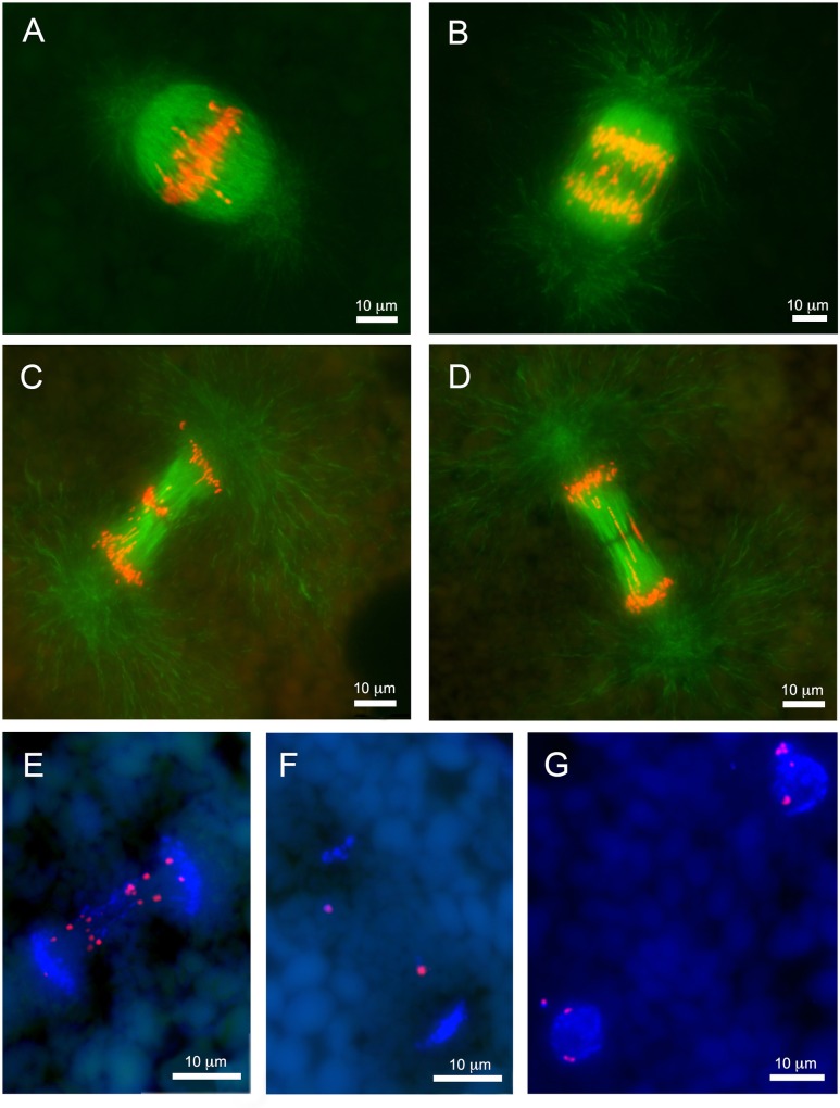
Fig 2. Lagging chromosomes are abundant during early stages of embryonic development.
(A-D) Images of paraffin sections from lamprey embryos at 1 dpf. Anti-beta-tubulin immunolabeling: (A) metaphase, (B) anaphase A, (C) anaphase B with conglomerated chromatin in the equatorial area, (D) anaphase B with longitudinally stretched lagging chromatin. (E-F) Fluorescence in situ hybridization of the Germ1 probe to embryo cells at 2 dpf from paraffin sections. (E) Anaphase with lagging chromosomes contain multiple signals for Germ1, somatic chromosomes retain a single pair of Germ1 signals. (F) Late anaphase with two Germ1-positive micronuclei situated between condensing daughter nuclei. (G) Interphase cells with a single pair of Germ1 signals in their main nuclei and additional signals in micronuclei.

