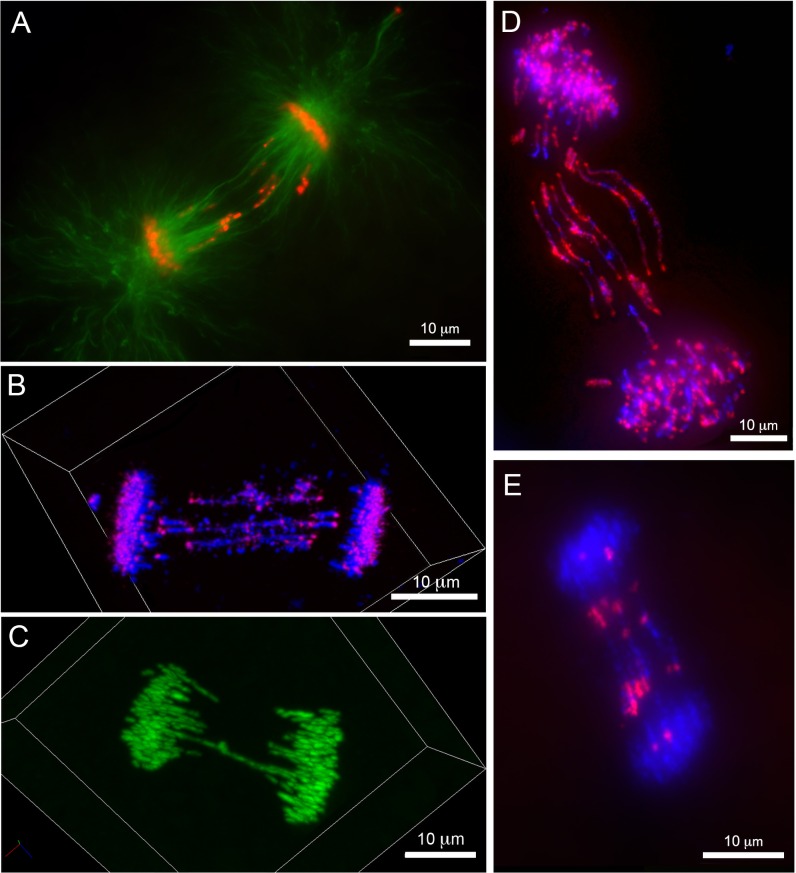
Fig 4. Morphology of anaphase lagging chromatin in intact embryonic cells.
Immunolabeling and hybridization of intact, PACT-cleared, embryos (2 dpf). (A) immunolabeling with anti-beta-tubulin, lagging chromatin is oriented along the polar microtubules. (B) Confocal image of an anaphase labeled with a centromere specific probe (Cot1 FISH). Centromeres of lagging chromosomes are oriented toward the poles of mitotic spindle. (C) Confocal image of an anaphase from an embryo at 1dpf: lagging chromosomes form equatorial contacts, bridging between the poles of the mitotic spindle. (D) An anaphase with multiple bridging chromosomes, hybridized with a probe to repetitive DNA (Cot2 FISH). Punctate signals corresponding to centromeres are oriented toward the spindle poles but lag behind retained chromosomes. Sites of apparent contact between sister chromatids hybridize strongly to Cot2 DNA, suggesting that repetitive DNA may participate in establishing these contacts. (E) Fluorescence in situ hybridization of the Germ1 probe to an anaphase with several bridging chromosomes. Germ1 signals are symmetrical, further supporting the interpretation that bridging features consist of pairs of sister chromatids, though notably, Germ1 signals do not appear to overlap with the zone of contact between sister chromatids.

