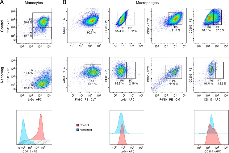
Fig 4. Representative dot plots and histograms of flow cytometric investigations of the effect of SPIO labelling on cell functionality.
(A) Unlabelled control (upper row) and Nanomag labelled luc+ monocytes (middle row) were stained against monocytic markers Ly6c and CD115. Upon iron particle labelling, monocytes reduced their expression levels for CD115, which is clearly visible in the depicted histogram for CD115 marker (lower row). (B) Luc+ MΦ without and with SPIO-labelling were tested for their expression levels of CD68, CD80, CD86, CD206 and F4/80. The monocytic markers Ly6c and CD115 served as negative controls. For both, labelled and unlabelled MΦ, F4/80-positive cells were also positive for CD68, CD80, CD86 and CD206. No change in expression levels of Ly6c could be detected upon particle uptake, whereas the CD115+ population of control cells minimized due to contrast agent incorporation. n = 2 for both cell types.

