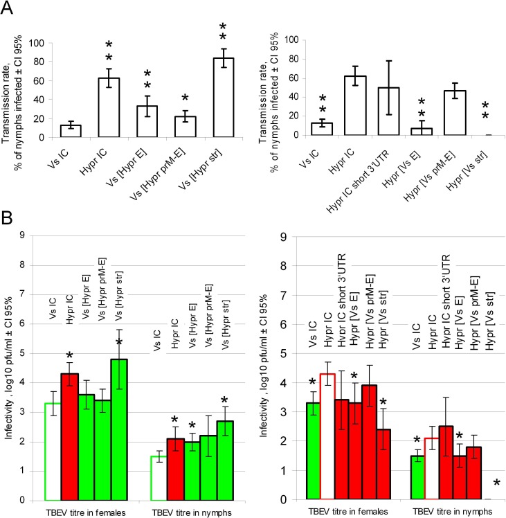
Fig 6. TBEV transmission and replication in I. ricinus ticks.
Each adult tick female was injected with 500 pfu of virus (as specified). After 14 days two adult females were co-fed with 15 nymphs for three days and virus titres in females and nymphs were determined by plaque infectivity assay. Left and right panels illustrate biological effects of gene replacements in Vs-based and Hypr-based chimaeras respectively. Mean values are shown with error bars indicating 95% confidence intervals. A) Transmission rate of TBEV between co-feeding ticks is presented as a proportion of nymphs that acquired TBEV infection after co-feeding with infected female ticks. Depending on levels of statistically significant differences (Mann-Whitney test) between chimaeric and control viruses, results are labelled with double (p < 0.01) or single asterisks (p < 0.05). B) Virus titres of TBEV in nymphs and females of I. ricinus. Vs- and Hypr-based chimaeras are identified as green and red bars respectively; the control viruses are shown as empty bars. Highly significant differences (p < 0.05, unpaired two-tailed t-test) between chimaeric and control viruses are labelled with an asterisk.

