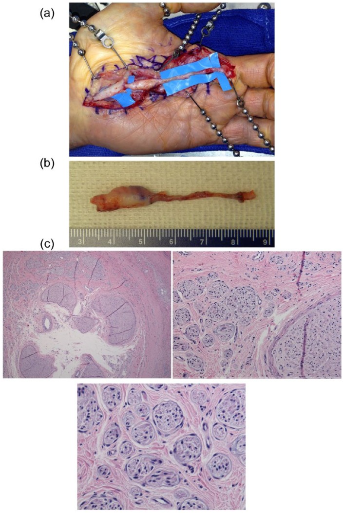
Figure 1.
(a) Exposure of the previously failed surgery reveals the allograft with a proximal neuroma of the common digital nerve. (b) Recalcitrant neuroma ex vivo. (c) A cross-section hematoxylin and eosin stain of neuroma. Low-power field demonstrating fascicles within the lumen of the allograft (upper left). High-power field magnification of fascicles varying in size, characteristic of neuromas (upper middle and lower).

