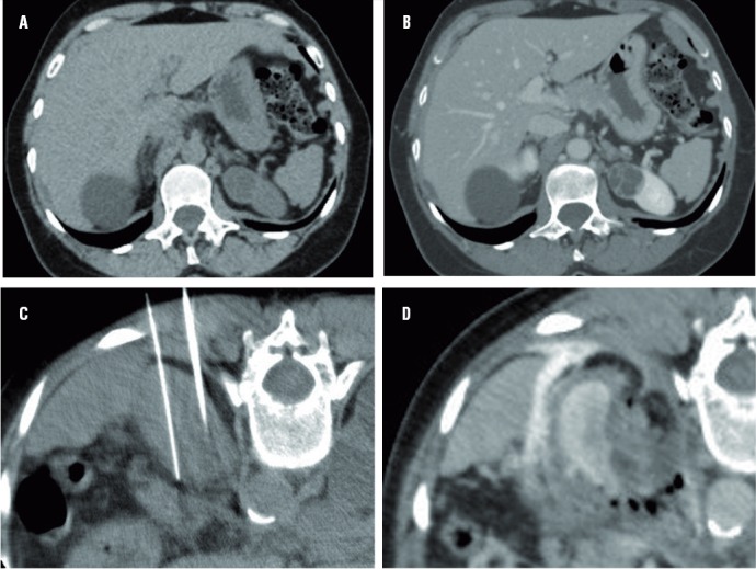
Figure 1. – (a-d) Tomographic images of radiofrequency ablation of Bosniak IV cystic lesion. CT without intravenous contrast (a) and post contrast (b) shows the hypoattenuation lesion in the left kidney and the exophytic cortical cyst in the upper third of the left kidney, containing thick internal septa that enhances after contrast. Non-enhanced CT (c) shows the positioning of the cluster needle in the axial plane. Enhanced-CT (d) immediately after the ablation shows the volumetric reduction of the lesion and the margins obtained after contrast injection, confirming proper treatment during the procedure.