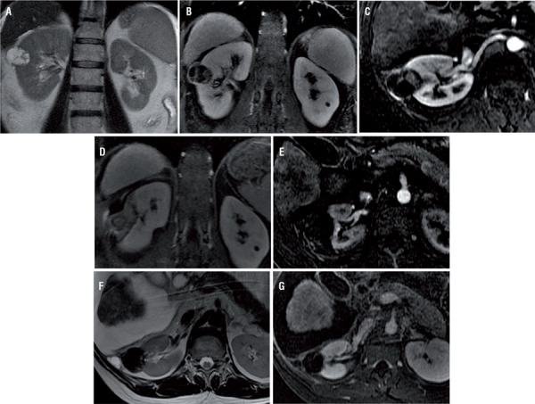
Figure 2. – (a-g) 48-years old man with a Bosniak IV lesion. Coronal T2-weighted (a), Coronal T1-weighted gradient echo axial MRI sequences with fat suppression (b) and coronal subtraction image (postcontrast arterial phase data - precontrast data) (c) show a cystic renal mass with thickened enhancing septa and a small solid component. Coronal T1-weighted gradient echo axial MRI sequences with fat suppression (d) performed immediately after the procedure show complete ablation of cystic lesion and no measurable enhancement within ablation zone. Axial subtraction image (postcontrast arterial phase data - precontrast data) (e) three months after the procedure show no enhancement and no recurrent of the neoplasm. Axial T2-weighted (f) and axial subtraction image (postcontrast arterial phase data - precontrast data) (g) 3 years after the procedure show ablation changes in the right kidney, without residual enhancement to suggest recurrent neoplasm.

