
Figure 1. Characterization of PCSK9 association with Lp(a).
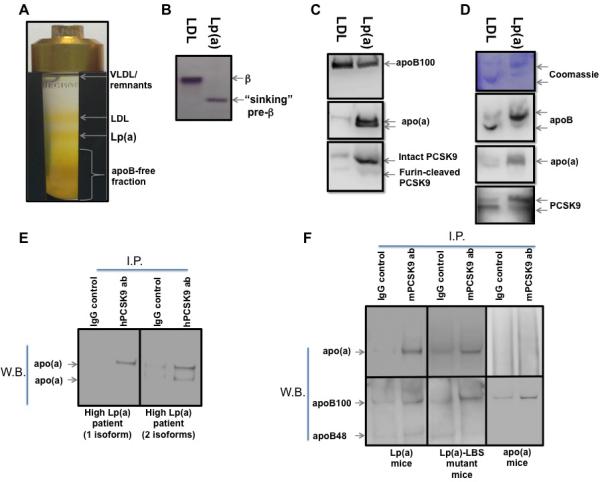
(A) lipoprotein isolation via iodixanol-based gradient ultracentrifugation. (B) agarose gel electrophoresis of LDL and Lp(a) fractions isolated as described in A, followed by neutral lipid staining. (C) Reducing gel immunoblot of LDL and Lp(a) fractions isolated as described in A. (D) Non-reducing PAGE of LDL and Lp(a) fractions isolated as described in A, followed by Coomassie protein staining (upper panel) and immunoblot (lower panel). (E) Immuno-precipitation of apo(a) using anti-PCSK9 antibody from plasma of subjects with one (left panel) or two (right panel) visible apo(a) isoforms. (F) Immuno-precipitation of apo(a) (upper panel) and apoB (lower panel) using anti-PCSK9 antibody from plasma of Lp(a) (left panel), Lp(a)-LBS mutant (middle panel), and apo(a) transgenic mice (right panel).