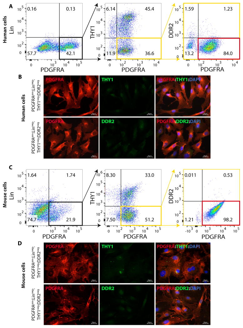
Figure 1. Isolation of FAPs from human and mouse hearts.
A, C. Flow cytometry plots showing the multi-step approach used to isolate FAPs from human (A) and mouse (C) hearts: non-myocyte fraction of cardiac cells were sorted to isolate cells positive for PDGFRA but negative for the hematopoietic progenitor markers CD32, CD11B, CD45, Lys76, Ly-6c and Ly6c, the stem cell and fibroblast marker THY1, and the fibroblast marker DDR2. Positive gates were set by analyzing signals from negative control samples, which were stained only with the corresponding IgG isotype for each marker. B, D. Immunofluorescence panels confirming the lack of expression of THY1 and DDR2 in human (B) and mouse (D) FACS isolated PDGFRApos:Linneg:THY1neg:DDR2neg cells.

