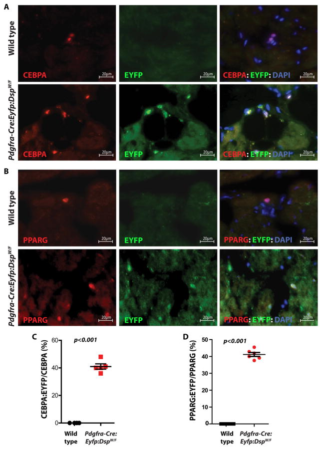
Figure 6. FAPs are a cell source of excess adipocytes in the heart of the Pdgfra-Cre:Eyfp:DspW/F lineage tracer mice.
A, B. Immunofluorescence panels showing co-expression of the reporter protein EYFP and the adipogenic markers CEBPA (A) and PPARG (B) in the myocardium of wild type and Pdgfra-Cre:Eyfp:DspW/F lineage tracer mice. Thin myocardial sections from wild type mice were included as controls. Approximately 41±4 % of cells expressing CEBPA also expressed EYFP in the heart of lineage tracer mice (C) (N=5 mice per group; 4 sections per mouse, 20 fields of 63X magnification per section). Similarly, 41±3 % of the cells that expressed PPARG also expressed EYFP (D) (N=6 mice per group; 4 sections per mouse, 20 fields of 63X magnification per section). The data indicate genetic labeling of the adipocytes by the Pdgfra locus in the heart of the lineage tracer mice.

