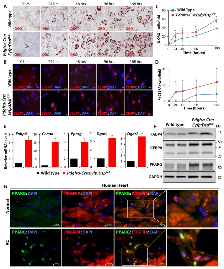
Figure 7. Enhanced adipogenesis in cardiac FAPs isolated from the DSP haplo-insufficient mice and detection of adipogenic FAPs in the human heart.
A, B. Oil Red O staining and CEBPA immunostaining showing accumulation of fat droplets (A) and expression of the adipogenic transcription factor CEBPA (B) in cardiac FAPs isolated from the Pdgfra-Cre:Eyfp:DspW/F lineage tracer mice, as compared to wild type mice, at 4 time points upon induction of adipogenesis with insulin, IBMX and DXM. C, D. quantitative data showing increased number of mature (ORO+) adipocytes (C) and CEBPA+ cells (D) in the cardiac FAPs from the transgenic mice as compared with wild type mice at each time point (N=3, ~300 cells counted at each time point for each group, *p<0.05). E. qPCR data for selected adipogenic genes after 4 days of adipogenesis induction showing marked increase in the transcript levels of Fabp4, Cebpa, Pparg, Dgat1, and Dgat2 in cardiac FAPs isolated from the Pdgfra-Cre:Eyfp: DspW/F mice as compared to wild type mice (N=3, * p<0.05). F. Immunoblots showing increased protein levels of adipogenic markers FABP4, CEBPA, and PPARG in cardiac FAPs isolated from the heart of lineage tracer mice after 4 days of adipogenic induction. G. Immunofluorescence stained panels from a control human heart and a human heart from a patients with arrhythmogenic cardiomyopathy (AC), showing co-expression of PDGFRA and the adipogenic transcription factor PPARG, suggestive of the presence of FAPs in transition to adipocytes in the human heart with AC.

