Fig. 1.

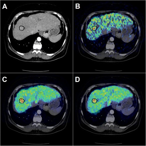
Contrast-enhanced CT image (arterial phase) with VOI-definition around the hypervascular HCC tumour (a) fused with the metabolic PET image (b), with the 1-h static image (c), and with the 2-h static image (d)

Contrast-enhanced CT image (arterial phase) with VOI-definition around the hypervascular HCC tumour (a) fused with the metabolic PET image (b), with the 1-h static image (c), and with the 2-h static image (d)