Fig. 2.

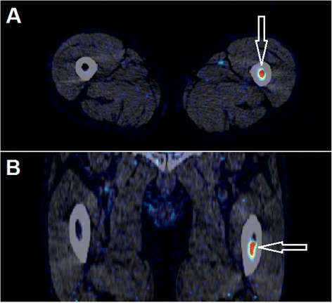
Arrows show a metastasis in the left femoral bone of patient #6 detected on a static 18F-FDGal PET image (visible on both static images; here, the 1-h static image is shown). a Transaxial slice. b Coronal slice

Arrows show a metastasis in the left femoral bone of patient #6 detected on a static 18F-FDGal PET image (visible on both static images; here, the 1-h static image is shown). a Transaxial slice. b Coronal slice