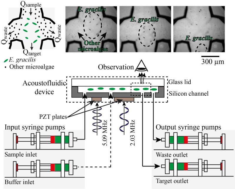
FIG. 2.
Evaluation of the on-chip acoustofluidic device. The flow rates were controlled by syringe pumps mounted with syringes connected to the inlets and outlets of the microchannel. The separated cells were enumerated by using an image-based cytometer. The observation of the device's functionality was carried out with a microscope with a high-speed camera at a frame rate of 1200 fps. As a proof-of-principle demonstration, a mixture of E. gracilis (50–100 μm sized) and M. aff. jurisii (5–10 μm sized) was infused into the sample inlet by one of the syringe pumps at a controlled flow rate of 500 μl/min. The sequence of the image frames that capture the dynamical process at the trifurcation-shaped junction in the figure indicates a successful demonstration of directing E. gracilis cells into the central channel and M. aff. jurisii cells and other small particles into the side channels.

