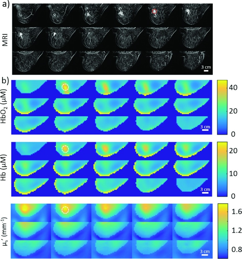
FIG. 7.
MRI and DOT images of the left breast of a 79-year-old post-menopausal Caucasian female with breast cancer, viewed as lateral to medial sagittal slices from the upper left. (a) Subtraction images from a DCE-MRI (Gadolinium). Tumor biopsy confirmed as invasive mammary carcinoma (ductal and lobular features) with intermediate grade nuclei. (b) 3D multispectral frequency-domain DOT reconstruction of the HbO2 and Hb concentrations (μM) as well as the at 785 nm. The approximate tumor location in the MRI, as well as the corresponding area in each DOT image, is indicated by a dashed circle of 30 mm in diameter. Note, MRI and DOT breast compressions are similar but are not identical. Interesting features are apparent in the images with high HbO2, Hb, and scattering near the tumor region (identified with biopsy clip in the MRI).

