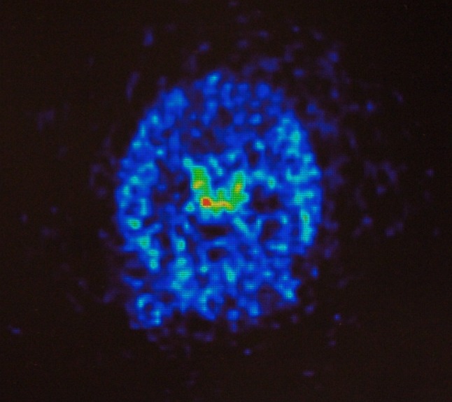
Fig. 9.

Positron emission tomography image, transverse section, of a living rat brain following perispinal extrathecal administration of 64Cu-DOTA-etanercept, imaged 5–10 min following the administration of etanercept. Note enhanced signal in the choroid plexus. Reproduced from Tobinick et al. [18]
