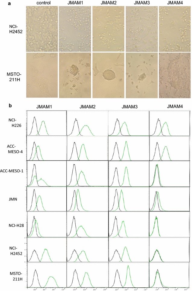
Fig. 1.

Reactivity of JMAM mAbs with mesothelioma cell lines. a Morphological changes by JMAM mAbs. NCI-H2452 cells were incubated with hybridoma supernatants for 72 h and observed using visible light microscopy. RPMI-1640 medium with 10 % FCS served as the control (upper column). These findings were also reproduced using MSTO-211H cells (lower column). b Flow cytometry analysis of JMAM mAb reactivity. Mesothelioma cell lines were incubated with the hybridoma supernatant (green histogram) or control mouse IgG (black histogram), subsequently stained with Alexa Flour®-488 labeled anti-mouse IgG Ab and analyzed using flow cytometry
