
Fig. 2.
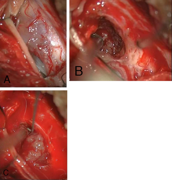
Intraoperative view of the spinal cord after opening the dura mater via dorsal laminectomy; the tumor shines brightly in (A). Melanocytoma with visible caudal tumor edge (B) and partially dissected melanocytoma (C).
Official websites use .gov
A
.gov website belongs to an official
government organization in the United States.
Secure .gov websites use HTTPS
A lock (
) or https:// means you've safely
connected to the .gov website. Share sensitive
information only on official, secure websites.
Intraoperative view of the spinal cord after opening the dura mater via dorsal laminectomy; the tumor shines brightly in (A). Melanocytoma with visible caudal tumor edge (B) and partially dissected melanocytoma (C).