
Fig. 4.
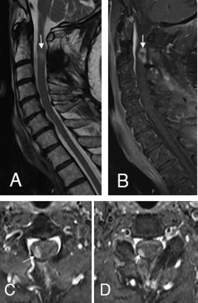
The followup MRI after 3 months indicates the solid enhancing tumor recurrence (B) at the right adherent lateral resection border at the C2 (C) and C3 level (D). The signs of cervical myelopathy were still recognizable on the sagittal T2-weighted (A) images.