
Fig. 2.
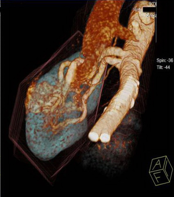
62-year-old male with renal arteriovenous malformation. Findings: Volume-rendered oblique coronal image of right kidney in arterial phase of CT angiography showing aneurysmal dilatation of segmental branch at lower pole with early opacification of renal vein suggestive of arteriovenous malformation. Technique: Scanner: Siemens Somatom sensation 64, 200Eff mAs, 120 kV, slice thickness 10mm, intravenous contrast: 100 ml Omnipaque 350 mg/dl.