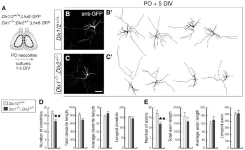
Figure 2. Interneurons from neocortex of Dlx1−/−;Dlx2+/− compound mutants develop neuronal processes with fewer branches.

(A) Experimental assay used to characterize the morphology of interneurons.
(B, C) Examples of interneurons isolated from P0 neocortex and immunostained with anti-GFP after 5 DIV. (B′, C′) are drawings of representative neurons.
(D, E) Quantification of axonal and dendritic morphology from control (gray bars) and Dlx1−/−;Dlx2+/− (black bars) interneurons after 5 DIV. Student’s t-test revealed significantly decreases in the numbers of dendritic and axonal branches in mutant cells (dendrites: mutant: 16.6 ± 1; control: 21.8 ± 1.2, p < 0.01; axons: mutant: 12 ± 0.8; control: 17.3 ± 1.6, p < 0.01; n = 30). ** p < 0.01.
Scale bar = 100 μm (B,C).
