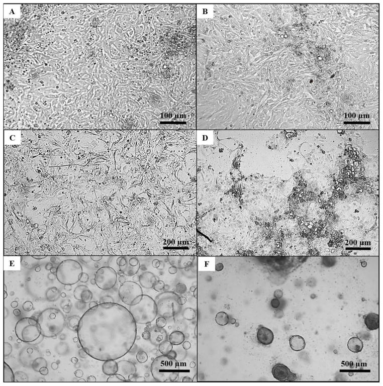
Figure 2. Light microscopy of human intestinal epithelial cultures.
A: A less differentiated monolayer of epithelial cells (Passage 4, Day 3). B: A more differentiated monolayer of epithelial cells (Passage 4, Day 3). C: A less differentiated monolayer of epithelial cells prior to transforming cells into a more differentiated monolayer of cells. D: A more differentiated monolayer generated from supporting monolayers with ENRYG-50Wnt3A and ENRY-DAPT-5Wnt3A. E: Spheroids (Passage 8, Day 5) generated from monolayers cells under light microscopy at 25x magnification. F: Enteroids (Passage 9, Day 16) generated by supporting spheroids with ENRYG-50Wnt3A and ENRY-DAPT-5Wnt3A under light microscopy at 25x magnification.

