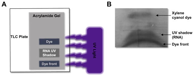
Fig. 4.
Visual representation of UV shadowing. (A) Schematic of set-up for UV-shadowing of RNA. The denaturing urea-PAGE gel (shown in grey) is positions on top of the TLC plate (shown in white). UV light (purple) is shined on top of the gel. A shadow will appear in the place where the RNA aptamer is on the gel. The RNA aptamer runs between the top dye (xylene cyanol) and the dye front (bromophenol blue). (B) UV shadow of an RNA aptamer visualized using the TLC plate method. The RNA is run on a denaturing Urea-PAGE gel until the dye front reaches the bottom of the gel. The RNA band (shadow) can be easily excised using a clean razor blade and the RNA eluted from the gel in 1X TE buffer.

