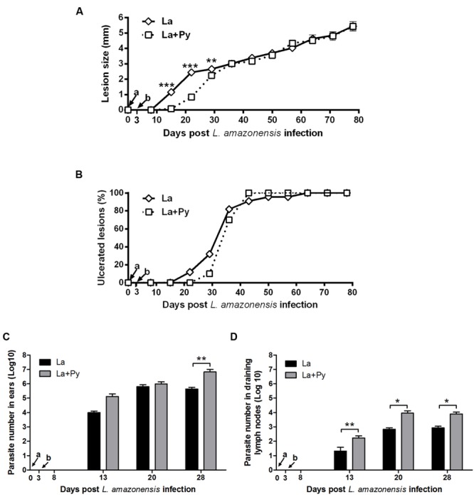
FIGURE 4.

Evaluation of L. amazonensis infection. (A) Lesion development, (B) onset of open ulcers, and (C) parasite quantitation in infected ears and (D) draining lymph nodes of BALB/c mice after intradermal infection in the ears with L. amazonensis promastigotes (La) or co-infected with L. amazonensis and P. yoelii (La+Py). Parasite loads were individually determined by a limited dilution assay. Results are expressed by the mean ± SEM of twelve ears and lymph nodes. The Mann–Whitney test was used to compare parasite loads in ears and draining lymph nodes. ∗p < 0.05 and ∗∗p < 0.01, respectively. Arrows indicate the time of L. braziliensis (a) and P. yoelii (b) infection.
