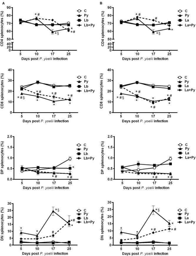
FIGURE 8.

Alterations in spleen T-cell subsets after infection with P. yoelii (Py), L. braziliensis (Lb), L. amazonensis (La), or co-infection with Leishmania sp. and P. yoelii. The phenotype of T subsets in the spleen were determined by flow cytometry at days 5, 10, 17, and 25 post P. yoelii infection. The percentages of CD4+, CD8+, CD4-CD8- double negatives (DN), and CD4+CD8+ double positive cells (DP) obtained in uninfected controls (open circles) were compared to those obtained in Py single-infected mice (black triangles) and in animals co-infected (black diamonds with dashed line) or single infected (black squares) with L. braziliensis. (A) or L. amazonensis promastigotes (B). Results are expressed by the mean ± SEM of 4–6 mice per group. The Mann–Whitney test was used to compare groups. (∗) statistically different from control, (#) statistically different from Leishmania sp. and (§) statistically different from Leishmania sp./P. yoelii co-infected group.
