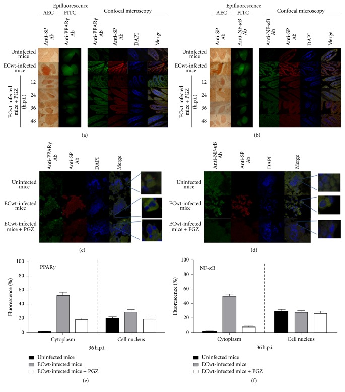
Figure 5.
Immunochemistry, immunofluorescence, and confocal microscopy analyses of PPARγ and NF-κB in villus cells from ECwt-infected mice treated with PGZ. (a) Histological cross sections of duodenal villi from ECwt-infected mice that had been treated with PGZ were analyzed by immunocytochemistry for the presence of ECwt SP (EAC) or epifluorescence for the presence of PPARγ (FITC). Histological cross sections were observed using confocal microscopy for the identification of PPARγ (green) and rotavirus SP (red). Nuclei were counterstained with DAPI (blue). Representative photographs are shown. (b) Histological cross sections analyzed for the presence of NF-κB and rotavirus SP as indicated in (a). (c) Villi isolated from ECwt-infected and noninfected mice that have been treated or not with PGZ were processed and analyzed at 36 h.p.i. for the subcellular location of PPARγ and ECwt SP using confocal microscopy. Representative micrographs are shown. (d) Villi isolated from ECwt-infected or noninfected mice were treated or not as described in (c), except that they were analyzed for the subcellular location of NF-κB and ECwt SP. Representative micrographs are shown. (e) Villi from ECwt-infected or noninfected mice (n = 3 per experiment) that had been treated or not with PGZ were subjected to confocal analysis for the quantification of subcellular location (nucleus or cytoplasm) of PPARγ. (f) Villi from ECwt-infected or noninfected mice were treated as described in (e), except that they were analyzed for the quantification of subcellular location (nucleus or cytoplasm) of NF-κB. Data are shown as mean ± SD from three independent experiments performed in duplicate.

