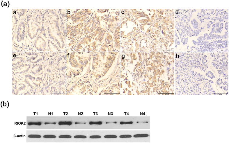
Figure 3. Representative patterns of RIOK2 and NOB1 immunohistochemical staining in NSCLC tissues as determined by IHC with TMA sections.
(a) a weak RIOK2 staining in well-differentiated NSCLC tissues, b positive RIOK2 staining in moderately differentiated NSCLC tissues, c strong positive RIOK2 staining in poorly differentiated NSCLC tissues, d absent RIOK2 staining in an isotype control, e weak NOB1 staining in well-differentiated NSCLC tissue, f positive NOB1 staining in moderately differentiated NSCLC tissue, g strong positive NOB1 staining in poorly differentiated NSCLC tissue, and h absent RIOK2 staining of in an isotype control (200×). (b) Protein expression of RIOK2 in the NSCLC patients as detected by western blotting of the fresh frozen tissue samples (T: NSCLC tumour tissue, N: paired adjacent normal lung tissue).

