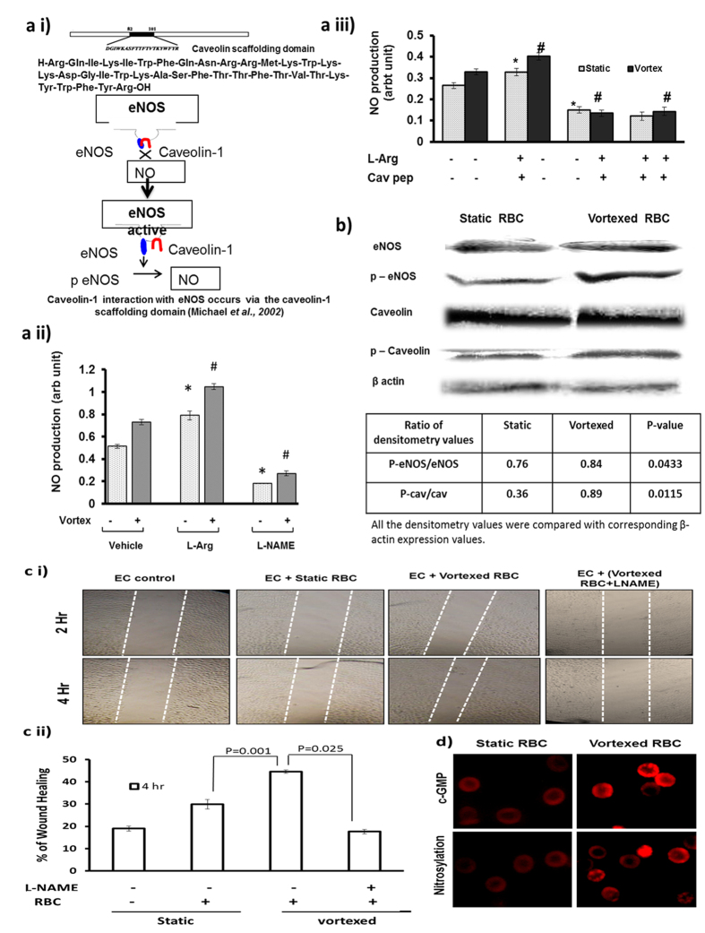
Figure 5. Production of NO by vortexed RBCs stimulates wound healing and is e-NOS dependent.
(a i) Representative image shows role of caveolin in eNOS activation. Static or vortexed RBCs were incubated with e-NOS substrate (L-Arginine), NOS inhibitor (L-NAME) or caveolin scaffold domain (82–101) inhibitor for 30 minutes at 37 °C, and NO level was measured using DAF-FM. (a ii) Bar graph shows NO production was significantly increased in static RBCs in presence of L-arginine (n = 3; *p = 0.007), and decreased in presence of L-NAME (n = 3; *p < 0.001). Similarly, (a iii) NO production was significantly increased in vortexed RBCs in presence of L-arginine (n = 3; **p < 0.001) and decreased in presence caveolin scaffolding peptide (n = 3; #p < 0.001). (b) Vortexing induced phosphorylation of RBC eNOS and caveolin-1. Static and vortexed RBC showed similar basal levels of eNOS and caveolin proteins in RBC ghosts. Vortexed RBCs showed increased levels of phosphorylated e-NOS (serine-1177) (p = 0.0433) and phosphorylated caveolin (Tyr-14) (p = 0.0115) compared with static RBC. (c) NO produced by vortexed RBC promotes wound healing in EAhy926 cells. Static and vortexed RBCs (3 × 10⁶/3 mL) were incubated with the wound created in EAhy926 monolayer for 30 minutes in a CO2 incubator at 37 °C. Distances migrated by wounded cells were imaged and measured at the 4h time point. (c i) Representative image at 0th and 4th hour was captured by IX71 Olympus fluorescence microscope using a 4X objective lens and NA 0.10. (c ii) Wounded cells treated with vortexed RBC showed 20% greater migration than wounded cells treated with static RBC (n = 3; *p = 0.001). (d) Immunofluorescence images of vortexed and static RBC show TRITC-positive spots stained by anti-cGMP and anti nitrocysteine antibodies. Images shows significant increased intensity of cGMP (n = 3; #p = 0.010; t-test) and nitrosylation (n = 3; *p < 0.001; t-test) in vortexed versus static RBC. Images were by OlympusIX71 microscope, 60× magnification and oil-immersion objective lenses with a numerical aperture (NA) of 1.42.

