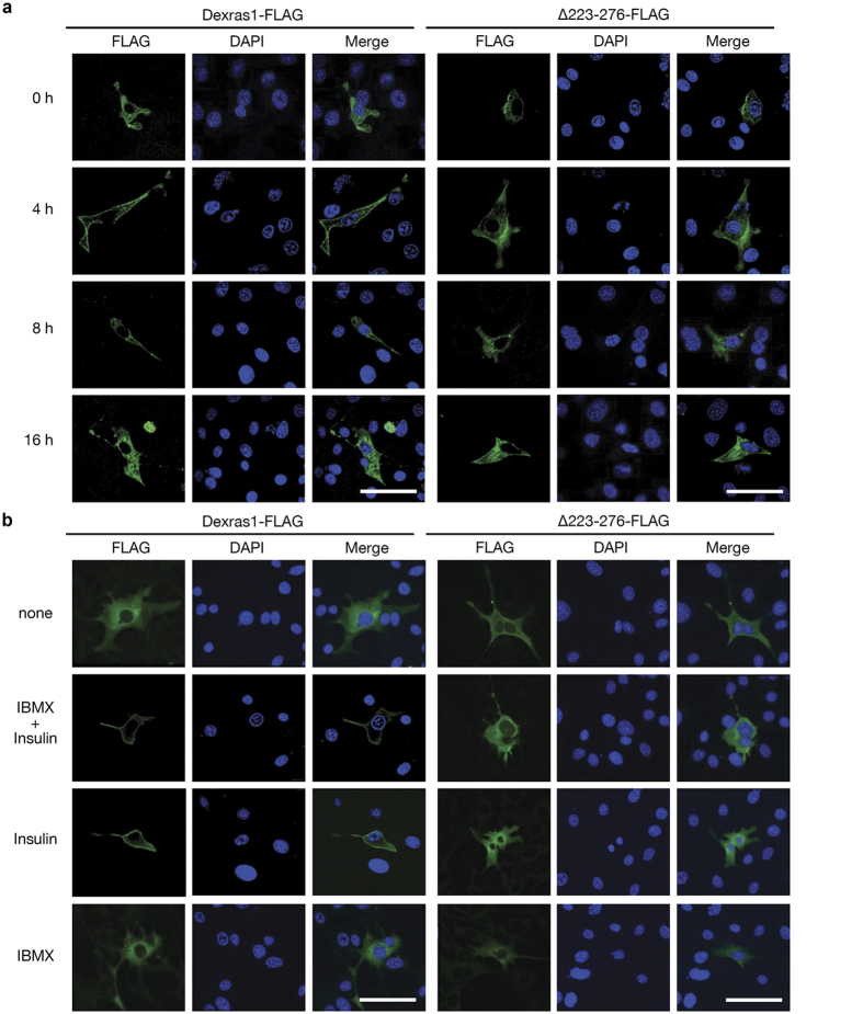
Figure 2. Dexras1 translocates to the plasma membrane in response to insulin signaling.
3T3-L1 cells were transfected with pcDNA3-Dexras1-FLAG or pcDNA3-∆223-276-FLAG vector. Cellular localization of Dexras1 was analyzed by confocal microscopy. Transfected cells were incubated with (a) IBMX, dexamethasone, and insulin for the times indicated, or (b) various differentiation cocktails for 1 h. To visualize Dexras1 protein, cells were fixed and subjected to immunofluorescence analysis with antibody against FLAG (green). Nuclei were stained with DAPI and fluorescence was visualized by confocal microscopy. Scale bar = 50 μm.

