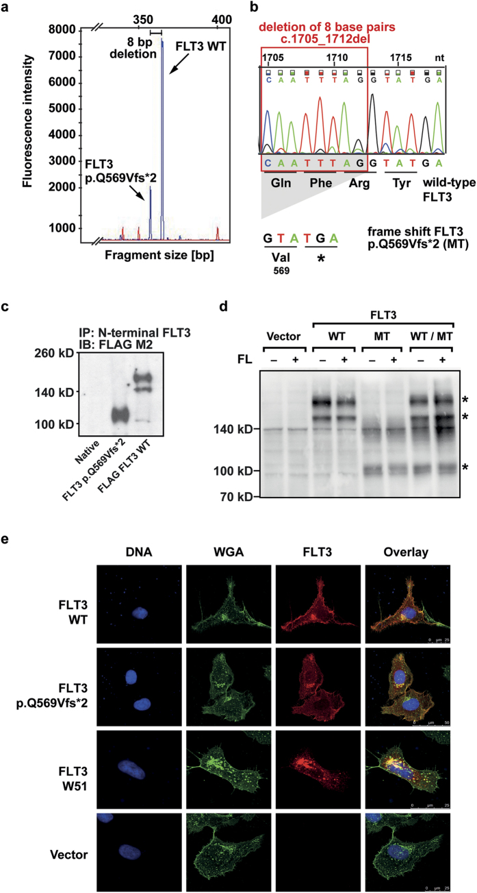
Figure 1. Identification of the mutation in the patient sample and expression of the truncated FLT3 p.Q569Vfs*2 receptor.
(a) Blast cells were isolated from the bone marrow of a relapsed AML patient. mRNA was isolated and reverse transcribed. The FLT3 cDNA was amplified and fragment analysis was performed. Arrows indicate fragments for FLT3 WT and FLT3 p.Q569Vfs*2. The peaks differ by eight base pairs in their fragment size. (b) Sanger sequencing revealed a deletion of eight nucleotides, leading to a frameshift and a premature stop codon within the FLT3 gene. The chromatogram is shown for the wild-type FLT3, nucleotide triplets and the corresponding amino acids are shown for the FLT3 WT and the frameshift mutation FLT3 p.Q569Vfs*2 sequence. (c) Phoenix eco cells were transfected with FLAG-tagged FLT3 WT and FLAG-tagged FLT3 p.Q569Vfs*2. After cell lysis the FLT3 protein was immunoprecipitated from whole cell lysates with an N-terminal FLT3 antibody (SF1.340). After blotting the FLAG-tagged FLT3 was detected with an FLAG M2 antibody. One representative experiment is shown. The blot was cropped to improve the clarity of the image. (d) Ba/F3 cells stably expressing the indicated constructs. After cell lysis the FLT3 protein was detected with an N-terminal FLT3 antibody (4B12). One representative experiment is shown. FLT3 bands are indicated by asterisks. The blot was cropped to improve the clarity of the image (MT = FLT3 p.Q569Vfs*2). (e) Immunofluorescence of FLT3 (red), glycoconjugates (green) and counterstaining of DNA (blue) in transiently transfected U2OS cells. One representative image of each construct is shown.

