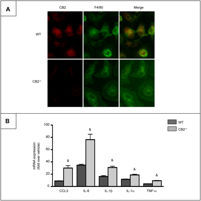
Figure 1. Macrophages from CB2Mye−/− mice display enhanced pro-inflammatory properties.
(A) Representative images of CB2 (red) and F4/80 (green) labeling in peritoneal macrophages isolated from WT and CB2Mye−/− mice (original magnification x400). (B) mRNA expression of CCL3, IL-6, IL-1β, IL-1α and TNF-α in Kupffer cells isolated from WT and CB2Mye−/− mice and exposed to 1 ng/ml of LPS for 6 hours. Data are mean ± SEM of 5–10 samples per condition. & p < 0.05 for WT vs CB2Mye−/− mice.

