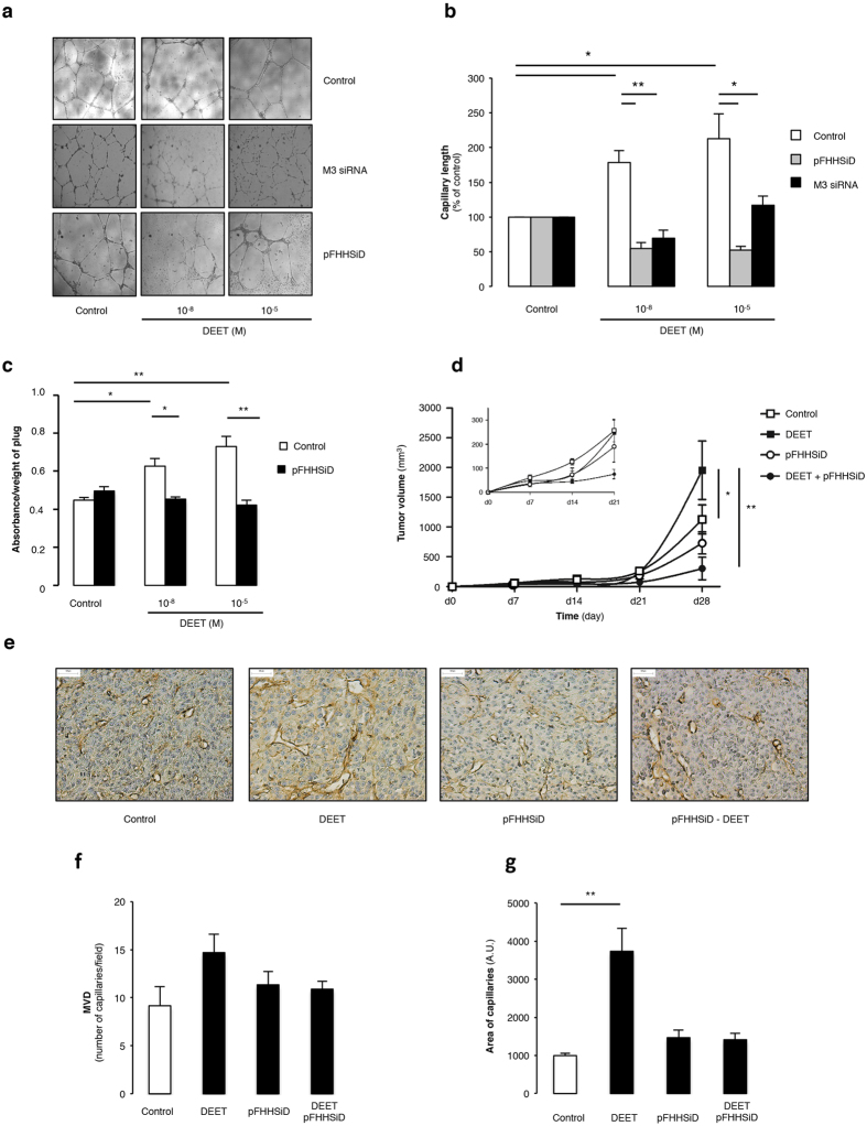
Figure 1. In vitro and in vivo pro-angiogenic properties of DEET.
(a) Representative illustrations of HUVEC on ECMgel, treated during 24 h in absence or presence of DEET 10−8 M or 10−5 M with or without pFHHSiD 10−7 M or silenced with specific M3 receptor siRNA. (b) DEET enhances in vitro capillary formation. Pharmacological blockade with pFHHSiD or silencing with specific siRNA against M3 muscarinic receptor were used. Reproducible data were obtained from four independent experiments. (c) DEET promotes in vivo neovascularization in ECMgel model. At day 14, quantitative measurement of hemoglobin was reported as absorbance (arbitray units)/weight of plugs. Hemoglobin content is increased in plugs containing mouse aortic-derived EC pretreated with DEET. Data were obtained from four independent experiments. Results are expressed at mean ± SEM. (d) DEET potentiates in vivo tumor growth. U87MG cells (106) were injected subcutaneously into the flank of six-week old female nude Swiss mice. Mice were chronically treated with 10−5 M DEET or its solvent (saline). The treatment was initiated the day following tumor cell injection and tumor dimensions were measured weekly. Pharmacological blockade with pFHHSiD prevents DEET-induced tumor growth. Data are expressed as tumor volume (mm3) and are presented as mean ± SEM (n = 6 per group). (e) DEET increases tumor-associated neovascularization. A representative paraffin section of tumor from each group (n = 6) is shown and reveals CD31 staining in brown. (f) Mean MVD (number of vessels/field) and mean area of capillaries (g) from tumors of mice treated with DEET and/or with pFHHSiD or saline are graphically represented. *p < 0.05; **p < 0.01 compared to control (Kruskal-Wallis with Bonferroni correction).

