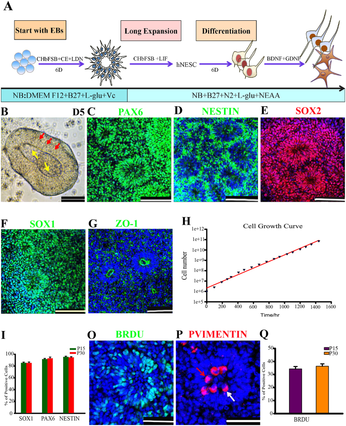
Figure 1. Generation of long-term self-renewal neuroepithelial stem cells.
(A) Schematic of the differentiation protocol for generating neuroepithelial stem cells (NESCs). CH, CHIR99021; bF, bFGF; CE, Compound E; NB, Neurobasal medium. The drawings were drawn by the author Xiaoqing Zhu. (B) Neural bodies at pdD5 attached and formed a two-layer structure after cultured onto laminin-coated plates for two additional days in the NESC culture media, yellow arrows and red arrows indicate two different layers, respectively. (C–G) Long-term cultured NESCs were organized into neural tube-like structures expressing neuroepithelial cell markers: PAX6, NESTIN, SOX2, SOX1 and ZO-1. (H) The growth curve of NESCs, displaying the prospect of exponential growth over serial passages. (I) Quantifications of PAX6, SOX1 and NESTIN positive cells in P15 (Passage 15) and P30 NESCs. Data expressed as mean ± s.d (n = 3) (P > 0.05 as Student’s t-test). (O) BrdU labeling identified most of the S-phase cells to be located on the basal surface of a neural tube (NT). (P) Mitotic marker phospho-VIMENTIN (p-VIMENTIN) positive cells were mainly distributed at the apical surface of a NT. The white arrow indicates symmetrical horizontal division orientation, and red indicates vertical division orientation. (Q) Quantification of BrdU incorporating cells in P15 and P30. Data expressed as mean ± s.d (n = 3) (P > 0.05 as Student’s t-test). Scale bars: 100 μm. Blue: DAPI, nuclear staining.

