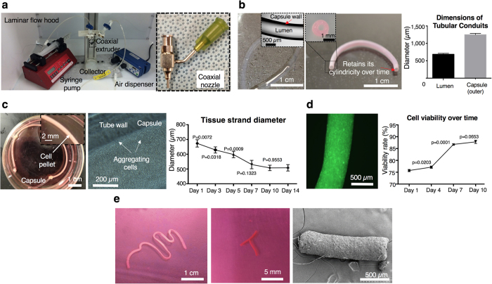
Figure 2. Fabrication of tissue strands.
(a) Left: Setup for co-axial extrusion of alginate capsules. Right: A home-made coaxial nozzle apparatus. (b) Left: Alginate capsules with well-defined tubular morphology. Middle: Capsules preserved their cylindrical morphology without any collapse in culture over time. Right: Controlled luminal and outer diameters (n = 6). (c) Left: Microinjected cell pellet inside alginate capsules supporting aggregation in a few days. Middle: Aggregation begins around the luminal surface of the capsule. Right: Tissue strand diameter decreases as aggregation progresses due to radial contraction and stabilizes after Day 10 (n = 6). (d) Left: Viable cells (green fluorescent) under confocal microscopic view. Right: Cell viability gradually increases over a 10-day culture period (n = 6). (e) Left: A nearly 8 cm long tissue strand after decrosslinking the capsule. Middle: Tissue strands were self-assembled into a “T” shape. Right: Well-defined cylindrical morphology was achieved as shown with SEM. All data are presented as average ± SD unless otherwise stated.

